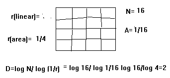
1. I. INTRODUCTION
his paper describes a computationally efficient technique to detect various texture characteristics as directional features in a given 3D digital image. The computational tool used for this purpose is '3D Rank Filters', which are essentially directional filters. These filters cause radical changes in the original content of a given image but precisely extract various textures.
Any given 3D MR image consists of texture features of tissues corresponding to muscle fibers in almost all directions. One can visualize major muscle fibers of a body component with naked eye. But most of the finer textures cannot be visualized even by an expert, in which case machine vision support system becomes quite handy. The algorithms presented in this paper could be used to detect texture patterns in al the three orthogonal axes of a 3D rectangular discrete coordinate system in which 3D digital image is displayed.
2. II. LITEERATURE SURVEY
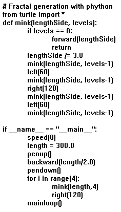
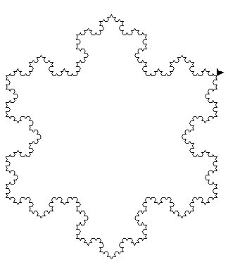
Apart from detecting latent textures in a given image, one can also artificially create texture images. Fig. 1 shows a 3D texture image, which is artificially generated using a cellular automaton rule. Two texture features are usually considered for image segmentation. They are (i) spatial frequency features and (ii) average gray level features. Either 'structural approach' or 'statistical approach' could be used for developing texture detection algorithms. Mostly statistical approach is considered for texture classification because of ease in parametrization and quantification of texture features.
Edge detection is a method by which one would be able to detect edge pixels details which help determine characteristics of texture complexities. For instance, directions of edges could be treated as characteristics of textures in determining patterns in the textures.
Consider a region with N pixels in a given image. Any gradient-based edge detector algorithm could be applied to this region, which would yield two outputs for every pixel p, viz, 'gradient magnitude Another technique to quantify texture is 'cooccurrence matrix', which defines features of a texture using certain spatial relations of similar gray values. Such numerical features could be used for texture classification. Some of the standard features from a normalized co-occurrence matrix are given below. where p[i, j] is the [i, j] th entry in a gray-level spatial dependence matrix, and Ng is the number of grayvalues in the quantized image. It is to be noted that the co-occurrence matrix based feature extraction will not yield comfortable visual perception.
3. III. PROPOSED METHOD
As outlined earlier, the term 'textures' refers to 'repeated patterns' in a given image. Consider the 27neighborhood window shown in Fig. 3. The cells 1, 2, 3, 4, 5, 6, 7, 8, 9 form the first plane, 10, 11, 12, 13, 14, 15, 16, 17, 18 the middle plane and cells 19, 20, 21, 22, 23, 24, 25, 26, 27 form the rear plane of the window. The given 3-D digital image is plane-wise raster-scanned by this window (See Fig. 3). In order to extract 3-D linear textures along an axis with a directional twist, one has to choose that particular axis and its associated rank of a particular directional twist. For example if one chooses the X axis and rank1 of zero directional twist, values in cells 2,11,20,23,26,17,8,5 would be read and stored in an array. The reading pattern is shown in Fig. 4. X-axis rank 2 consists of cells 11, 20, 23, 26, 17, 8, 5, 2 and the corresponding plane is perpendicular to X axis as given in Fig. 4 but with a directional twist of 45 degrees. One can construct four ranks in X-axis, four in Y-axis and another four in Z-axis as shown in Table 1. A total of 12 rank filters could be constructed in three axes which are called "3D Orthogonal Rank Filters".
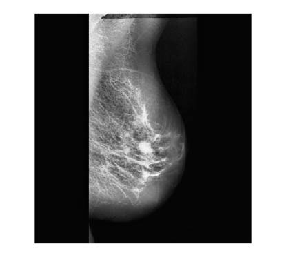
4. IV. TEXTURE CLASSIFICATION OF 3D MEDICAL IMAGES
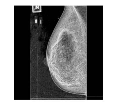
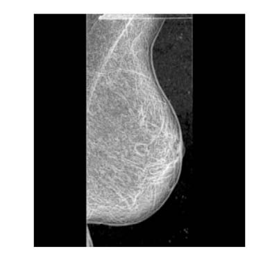
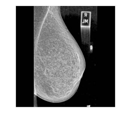
Textures of a medical image play an important role in support of a surgeon to decide the angle at which the surgical blade should be used to make incision so that the loss of blood due to surgery is kept minimum. A case study was carried out to verify the validity of the algorithm and the result of the study presented in Fig. 5, which is self-explanatory.
( ) All four texture versions of the image obtained using rank filters could be seen to provide a visual proof of the fact textures in an image are direction sensitive and so they could be used for image segmentation purposes.
Year 2021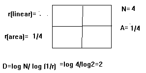
| Axes | Ranks | Cell sequences |
| X1 | 2,11,20,23,26,17,8,5 | |
| X | X2 X3 | 11,20,23,26,17,8,5,2 20,23,26,17,8,5, 2,11 |
| X4 | 23,26,17,8,5, 2,11,20 | |
| Y1 | 4,13,22,23,24,15,6,5 | |
| Y | Y2 Y3 | 13,22,23,24,15,6,5,4 22,23,24,15,6,5,4,13 |
| Y4 | 23,24,15,6,5,4,13,22 |
| Original 3D image statistics | |
| Pixels Count | 568089 |
| Pixels without black | 449360 |
| Red Min | 0 |
| Red Max | 252 |
| Red Mean | 90.0212677943069 |
| Red Standard Deviation | 65.4300367403954 |
| Red Median | 92 |
| Red Total Count | 568089 |
| Green Min | 0 |
| Green Max | 249 |
| Green Mean | 48.2296154299766 |
| Green Standard Deviation 56.6653938791041 | |
| Green Median | 33 |
| Green Total Count | 568089 |
| Blue Min | 0 |
| Blue Max | 249 |
| Blue Mean | 48.2296154299766 |
| Blue Standard Deviation 56.6653938791041 | |
| Blue Median | 33 |
| Blue Total Count | 568089 |
| Saturation Min | 0 |
| Saturation Max | 1 |
| Saturation Mean | 0.347210377454758 |