1. Introduction
2. RI Stands for Magnetic Resonance Imaging; once called Nuclear Magnetic Resonance
Imaging. The "Nuclear" was dropped off about 15 years ago because of fears that people would think there was something radioactive involved, which there is not. MRI is a way of getting pictures of various parts of your body without the use of x-rays, unlike regular x-rays pictures and CAT scans. A MRI scanner consists of a large and very strong magnet in which the patient lies. A radio wave antenna is used to send signals to the body and then receive signals back. These returning signals are converted into pictures by a computer attached to the scanner. Pictures of almost any part of your body can be obtained at almost any particular angle.
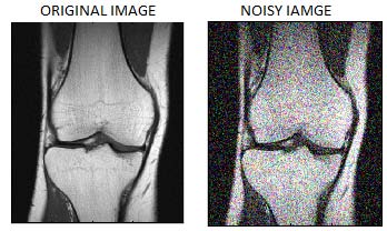
Medical information, acquired from MRI and composed of clinical data, images and other E-mail : [email protected] E-mail : [email protected] E-mail : [email protected] E-mail : rathore [email protected] Emission Computed Tomography (SPECT) and Positron Emission Tomography (PET). All these methods generate good quality of medical image [1] and each has its own specific features corresponding to the physical and physiological phenomena studied, as shown in "Fig. textured, or snowy appearance. Image noise comes from a variety of sources. No imaging method is free of noise, but noise is much more prevalent in certain types of imaging procedures than in others. Noise is also significant in MRI (Medical Resonance Imaging), CT, and ultrasound imaging. In comparison to these, radiography produces images with the least noise. Fluoroscopic images are slightly noisier than radiographic images. The presence of noise degrades the image quality and decreases visibility of lower contrast image. So there is a need for noise removal technique to improve the image quality and to recover the fine details of image which is required for perfect diagnostic. This paper is divided into seven sections. Section one gives idea about MRI and denoising. Section two shows a literature survey .Section three defines implementation of algorithm .Section four and five gives idea about Gaussian blur and anisotropic diffusion. Section six defines pproposed algorithm for denoising while section seven is conclusion II.
3. RELATED WORK
Various algorithms for image denoising are discussed in [2]. The de-noising of Magnetic Resonance Images using wave atom shrinkage is proposed in [3] and also proved that this approach achieves a better SNR compared to wavelet and curvelet shrinkages. A NL-Denoising method for Rician noise reduction is proposed in [4 & 5].In [6], Total Variation Wavelet-Based technique is used to remove a noise from MR image. The method to improve image quality using adaptive threshold based on contourlet transform is given in [7]. A new filter to reduce random noise in multicomponent MR images by spatially averaging similar pixels and a local principal component analysis decomposition using information from all available image components to perform the denoising process is proposed in [8]. An estimator using a priori information for devising a single dimensional noise cancellation for the variance of the thermal noise in magnetic resonance imaging (MRI) systems called ML estimator has been proposed in [9]. A noise removal technique using 4th order PDE is introduced in [10] to reduce noise in MRI images.. A phase error estimation scheme based on iteratively applying a series of non-linear filters each used to modify the estimate into greater agreement with one piece of knowledge, until the output converges to a stable estimate is introduced in [11].
III. IMPLEMENTATION Fig. 3 shows the block diagram, gives general idea for MRI denoising using intensity averaging method.
4. Fig. 3 : Block diagram of intensity averaging algorithm
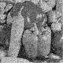
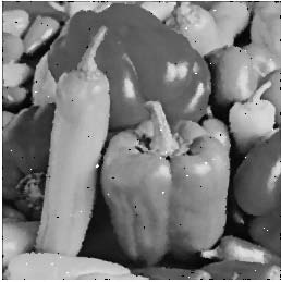
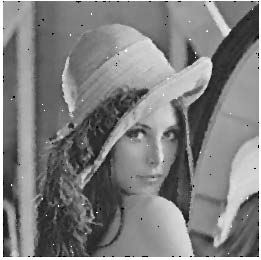
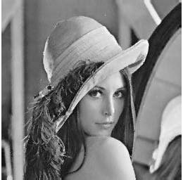
In proposed algorithm we have taken the image of [fig. 2] for evaluating our method. First we will apply amplitude correction on noisy MR image by finding forward and backward difference of intensity of pixels in X and Y direction. This gives average type of value to each pixel and then image is blurred by Gaussian filter and convolution. After completion of this amplitude correction, we apply a phase correction algorithm. Here, we are splitting amplitude corrected image into its red, green and blue band and then we are rotating each band by appropriate amount to correct the phase of MR image. After this, we are applying anisotropic diffusion and FFT to remove the noise from image.
5. GAUSSIAN BLUR
Gaussian blur is also known as Gaussian smoothing used to blur (smooth) the image. Typically it is used to reduce a random noise from the image. Mathemetically, Gaussian blur is equivalent to applying a convolution between image and Gaussian function [12,13]. Gaussian distribution in 1-D is given as,
??(??) = 1 ?2?? ?? ? ?? 2 2?? 2 ??(??, ??) = 1 2???? 2 ?? ? ?? 2 +?? 2 2?? 2Here, we are producing a discrete approximation of the Gaussian function before we perform the convolution as image is considered as a collection of pixels. Ideally we require an infinitely large convolution kernel because the Gaussian distribution is non-zero everywhere, but in practice we can truncate the kernel as Gaussian distribution in it is effectively zero, more than about three standard deviations from the mean. The degree of smoothing depends on the value of standard deviation. The Gaussian outputs a `weighted average' of each pixel's neighborhood, with the average weighted towards the value of the central pixels. This problem can be solved by anisotropic diffusion as discussed below.
V.
6. IMAGE FUSION AND ANISOTROPIC DIFFUSION
Image fusion describes the concept of combining multiple images into one image which gives more information compared to individual one [15]. Linear diffusion provides over smoothing of image as shown in fig. 3, we will use non-linear smoothing in which each pixel is treated with varying intensity depending on its neighboring value. In general, if (x,y) is a part of an edge ? apply little smoothing if not a part of an edge ? apply full smoothing This idea can be implemented by using a gradient function as given below.
????????(??) = ? ?? ???? , ?? ???? ?So non linear smoothing gives good intraregion smoothing as well as doesn't do much with interregion smoothing (edges and lines) as shown in fig. 4 The matter in an image is not heat, but brightness level. So, an image could be generalized to be a surface, where bright spots are "hot" and dark spots are "cold". So the idea is to use a varying size of
7. CONCLUSION
From the above result we conclude that, our algorithm is efficiently removing the noise from MR image. As number of iterations increases ("A"), we are getting more and more improved image. Experimental results show that, we are getting good Result in terms of PSNR and image quality. This algorithm is capable of removing noise from images and at the same time it is preserving fine details of images too. We also conclude that, for large value of iteration (A>25), increment in PSNR is less compared to small values of iterations (A<25).


![Fig. 4 : (a) Original image (b) image after linear smoothing (c) image after Non-linear smoothing This problem can be solved with anisotropic diffusion [17] when equation no.1 can be viewed as heat equation as shown below, It = ?I = (Ixx,Iyy)](https://computerresearch.org/index.php/computer/article/download/588/version/100522/3-Performance-Analysis-of-Intensity-Averaging_html/7968/image-7.png)
| Psnr2=25.98 | Mse2=15.51 |
| Psnr3=28.35 | Mse3=17.86 |
| Psnr4=24.37 | Mse4=17.95 |
| Psnr5=28.81 | Mse5=18.2 |
| Psnr2=27.10 | Mse2=16.63 |
| Psnr3=28.53 | Mse3=18.06 |
| Psnr4=27.60 | Mse4=18.07 |
| Psnr5=28.73 | Mse5=18.25 |
| Psnr2=27.75 | Mse2=17.28 |
| Psnr3=28.69 | Mse3=18.22 |
| Psnr4=28.70 | Mse4=18.23 |
| Psnr5=28.75 | Mse5=18.29 |
| VII. |